Diagnóstico endoscópico de Metaplasia Intestinal Gástrica en pacientes con Helicobacter pylori mediante endoscopia de imagen mejorada BLI
Resumen
Introducción: El Helicobacter pylori (H pylori) induce una potente respuesta inflamatoria por parte de la mucosa gástrica que produce una serie de alteraciones celulares secuenciales conocidas como la “cascada de Correa”: gastritis, atrofia, metaplasia intestinal (MIG) y, eventualmente, displasia y Cáncer Gástrico (CG). Al ser la MIG un marcador fenotípico temprano en la secuencia carcinogénica gástrica, queda identificado como un factor de riesgo para el CG, esto hace que el diagnóstico endoscópico de la MIG sea importante en pacientes con H pylori. Con el uso de la endoscopia con imagen mejorada (IEE) y Cromoendoscopia Electrónica BLI se puede hacer el diagnóstico de MIG, siendo los hallazgos en los patrones de superficie y vasculares muy consistentes. Objetivo: determinar la frecuencia de MIG Completa (MIGC) o MIG Incompleta (MIGI) en paciente con infección por H pylori mediante el diagnóstico endoscópico con IEE y BLI. Materiales y métodos: Se le realizó gastroscopias a 750 y 219 (38,8%) tenían diagnóstico histológico de infección por H pylori de los cuales 60 pacientes (27,4%) tenían diagnóstico endoscópico de MIG: MIGC 49 (82%) y MIGI 11 (18%). Diagnóstico histológico: MIGC 46 (77%) y MIGI 11 (18%). Conclusión: La IEE y BLI mostraron una alta sensibilidad (100%) y especificidad (97%) para la detección de MIG, lo que la convierte en una herramienta diagnóstica confiable.
ESCUCHE EL RESUMEN HANCIENDO CLIC EN EL IDIOMA DE SU PREFERENCIA:
EspañolEnglish
Portugues
Japones
Palabras clave
Texto completo:
PDFReferencias
Sugano K, Tack J, Kuipers EJ, Graham DY, El-Omar EM, Miura S, et al. Kyoto global consensus report on Helicobacter pylori gastritis. Gut. 2015;64(9):1353–67.
IARC Working Group on the Evaluation of Carcinogenic Risks to Humans. Schistosomes, liver flukes and Helicobacter pylori. IARC Monogr Eval Carcinog Risks Hum. 1994;61:177–240.
IARC Working Group on the Evaluation of Carcinogenic Risks to Humans. A review of human carcinogens: biological agents. IARC Monogr Eval Carcinog Risks Hum. 2009;100B:385–435.
Correa P. Human gastric carcinogenesis: a multistep and multifactorial process. Cancer Res. 1992;52(24):6735–40.
De Falco M, Manente L, Lucariello A, Esposito V. Molecular mechanisms of Helicobacter pylori pathogenesis. J Cell Physiol. 2015;230(8):1702–7. doi:10.1002/jcp.24933.
Uemura N, Okamoto S, Yamamoto S, Matsumura N, Yamaguchi S, Yamakido M, et al. Helicobacter pylori infection and the development of gastric cancer. N Engl J Med. 2001;345:784–9.
Filipe MI, Potet F, Bogomoletz WV, Whitehead R, Guillem G, Spiro M, et al. Incomplete intestinal metaplasia with sulfomucin in gastric cancer. Preliminary data from a prospective three-centre study. Gut. 1985;26(12):1319–26.
Rokkas T, Filipe MI, Sladen GE, Kaye P, Dixon MF. Detection of an increased incidence of early gastric cancer in patients with type III intestinal metaplasia who are closely followed. Gut. 1991;32(10):1110–3.
Bang BW, Kim HG. Endoscopic classification of intestinal metaplasia. Korean J Helicobacter Up Gastrointest Res. 2013;13:78–83.
Kaminishi M, Yamaguchi H, Nomura S, Oohara T, Sakai S, Fukutomi H, et al. Endoscopic classification of chronic gastritis based on a pilot study conducted by the Gastritis Research Society. Dig Endosc. 2002;14:138–51.
East JE, Vleugels JL, Roelandt P, Bhandari P, Bisschops R, Dekker E, et al. Advanced endoscopic imaging: European Society of Gastrointestinal Endoscopy (ESGE) Technology Review. Endoscopy. 2016;48(11):1029–45.
Barbeiro S, Libânio D, Castro R, Pimentel-Nunes P, Dinis-Ribeiro M. Narrow-band imaging: clinical application in gastrointestinal endoscopy. GE Port J Gastroenterol. 2018;26:40–53.
Dias-Silva D, Pimentel-Nunes P, Magalhães J, Libânio D, Dinis-Ribeiro M. The learning curve of narrow-band imaging in the diagnosis of precancerous gastric lesions using web-based video. Gastrointest Endosc. 2014;79(6):910–20.
Osawa H, Miura Y, Takezawa T, Harada R, Ino Y, Yoshizawa M, et al. Linked color imaging and blue laser imaging for the detection of the upper gastrointestinal tract. Clin Endosc. 2018;51(6):513–26.
Dohi O, Yagi N, Majima A, Onozawa Y, Itoh Y, Naito Y, et al. Diagnostic ability of magnifying endoscopy with blue laser imaging for early gastric cancer: a prospective study. Gastric Cancer. 2017;20(2):297–303.
Chen H, Liu Y, Lu Y, Lu J, Wang Y, Jin X, et al. Ability of blue laser imaging with magnifying endoscopy for the diagnosis of gastric intestinal metaplasia. Lasers Med Sci. 2018;33(8):1757–62.
Tahara T, Shibata T, Nakamura M, Yamashita H, Yoshioka D, Okubo M, et al. Gastric mucosal pattern by using magnifying narrow-band imaging endoscopy clearly distinguishes histological and serological severity of chronic gastritis. Gastrointest Endosc. 2009;70(2):246–53.
An JK, Song GA, Kim GH, Park DY, Kim DH, Lee BE, et al. Marginal turbid band and light blue crest, signs observed in magnifying narrow-band imaging endoscopy, are indicative of gastric intestinal metaplasia. BMC Gastroenterol. 2012;12:169.
Bansal A, Ulusarac O, Mathur S, Sharma P. Correlation between narrow-band imaging and nonneoplastic gastric pathology: a pilot feasibility trial. Gastrointest Endosc. 2008;67(2):210–6.
Liu H, Wu J, Lin XC, Zhou X, Li Y, Lin Q. Evaluating the diagnoses of gastric antral lesions using magnifying endoscopy with narrow-band imaging in a Chinese population. Dig Dis Sci. 2014;59(7):1513–9.
Ang TL, Pittayanon R, Lau JY, Ho KY, Rerknimitr R, Cheng YF, et al. A multicenter randomized comparison between high-definition white light endoscopy and narrow band imaging for detection of gastric lesions. Eur J Gastroenterol Hepatol. 2015;27(12):1473–8.
Lage J, Pimentel-Nunes P, Figueiredo PC, Libânio D, Dinis-Ribeiro M. Light-NBI to identify high-risk phenotypes for gastric adenocarcinoma: do we still need biopsies? Scand J Gastroenterol. 2016;51(5):501–6.
Pimentel-Nunes P, Libânio D, Lage J, Abrantes D, Coimbra M, Esposito G, et al. A multicenter prospective study of the real-time use of narrow-band imaging in the diagnosis of premalignant gastric conditions and lesions. Endoscopy. 2016;48(8):723–30.
Kanemitsu T, Yao K, Nagahama T, Hisabe T, Takaki Y, Hino S, et al. Extending magnifying NBI diagnosis of intestinal metaplasia in the stomach: the white opaque substance marker. Endoscopy. 2017;49(6):529–35.
Sha J, Wang P, Zhu B, Yang Y, Chen C, Yu M, et al. Acetic acid enhanced narrow-band imaging for the diagnosis of gastric intestinal metaplasia. PLoS One. 2017;12(2):e0170957
Tahara T, Tahara S, Tuskamoto T, Okubo M, Horiguchi N, Ishizuka T, et al. Magnifying NBI patterns of gastric mucosa after Helicobacter pylori eradication and its potential link to the gastric cancer risk. Dig Dis Sci. 2017;62(9):2421–7
Uedo N, Ishihara R, Iishi H, Yamamoto S, Yamamoto S, Yamada T, et al. A new method of diagnosing gastric intestinal metaplasia: narrow-band imaging with magnifying endoscopy. Endoscopy. 2006;38(8):819–24
Lim LG, Yeoh KG, Srivastava S, Khor CJ, Teh M, So JB, et al. Comparison of probe-based confocal endomicroscopy with virtual chromoendoscopy and white-light endoscopy for diagnosis of gastric intestinal metaplasia. Surg Endosc. 2013;27(12):4649–55
Rodríguez MT, Esposito G, Libânio D, Pimentel P, Dinis M. Image-enhanced endoscopy for gastric preneoplastic conditions and neoplastic lesions: a systematic review and meta-analysis. Endoscopy. 2020;52(12):1048–65
Dore MP, Lu H, Graham DY. Role of H. pylori eradication in gastric cancer prevention. Aliment Pharmacol Ther. 2022;55(Suppl 1):S14–21.
Shah SC, Piazuelo MB, Kuipers EJ, Li D. AGA clinical practice update on the diagnosis and management of atrophic gastritis: expert review. Gastroenterology. 2021;161(4):1325–32.
DOI: http://dx.doi.org/10.61155/gen.v79i3.751
IMÁGENES GEN
Autor: Dra. MARIA ISABEL RAMIREZ LÓPEZ - Volumen 79 (4) GEN 2025
 | |
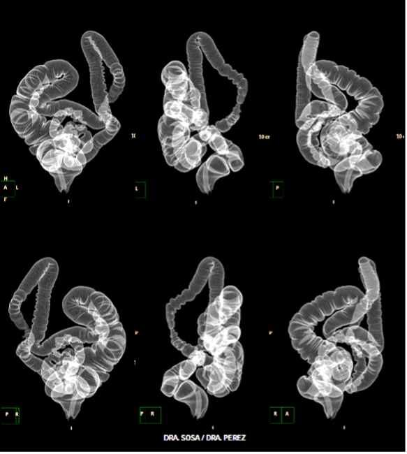
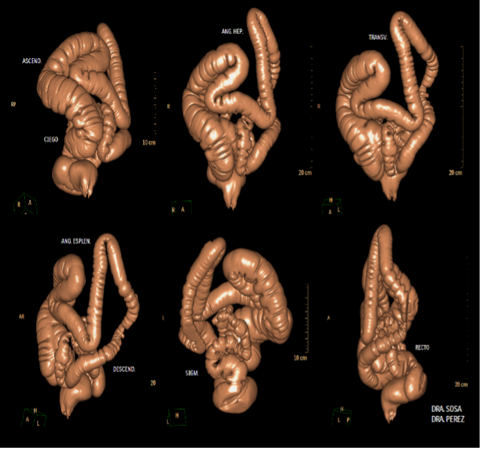
| POSPROCESAMIENTO DE IMÁGENES TOMOGRÁFICAS DE COLON PARA DIAGNOSTICAR CASOS DESAFIANTES. Autor: Dr. SERGIO MARTINEZ-MILLAN - Volumen 79 (4) GEN 2025 | |
 |  |
 |  |  |
ISSN: 0016-3503 e-ISSN: 2477-975X


