EXPERIENCIA CON CROMOENDOSCOPIA VIRTUAL COMPUTARIZADA (FICE) PARA LA DESCRIPCION Y CARACTERIZACION DE LESIONES COLORECTALES
Resumen
Palabras clave
Texto completo:
PDFReferencias
Kudo S, Hirota S, Nakajima T et al. Colorectal tumours and pit pattern. JClin Pathol 1994; 47: 880-885.
Sano Y, et al. Efficacy of magnifying chromoendoscopy for the differencial diagnosis of colorectal lesions. Digestive Endoscopy (2005) 17, 105-116.
Pohl J et al. Computed virtual chromoendoscopy: a new tool for enhancing tissue surface structures. Endoscopy 2007; 39: 80-83.
Kumagai Y, Inoue H, Nagai K et al. Magnying endosocpy, stereoscopic microscopy and the microvascular architecture of superfi cial esophageal carcinoma. Endoscopy 2002; 34: 369-375.
Yagi K, Nakamura A, Sekine A. Comparison between magnifying endoscopy and histological, culture and urease test fi ndings from the gastric mucosa of the corpus. Endoscopy 2002; 34: 376-381.
Fennerty MB. Tissue staining (chromoscopy) of the gastrointestinal tract. Can J Gastroenterol 1999; 13: 423-429.
Gono K, Yamaguchi M, Ohyama N. Improvement of image quality of the electroendoscopy by narrowing spectral shapes of observation light. Proc Int Congress Imaging Sci 2002; 5: 399-400.
Gono K, Yamazaki K, Doguchi N et al. Endoscopic observation of tissue by narrow−band illumination. Opt Rev 2003; 10: 1-5.
Sano Y, Kobayashi M, Hamamoto Y et al. New diagnostic method based on color imaging using narrow−band imaging (NBI) system for gastrointestinal tract. Gastrointest Endosc 2001; 53: AB125±.
Gono K, Obi T, Yamaguchi M et al. Appearance of enhanced tissue features in narrow−band endoscopic imaging. J Biomed Opt 2004; 9: 568-577.
Machida H, Sano Y, Hamamoto Y et al. Narrow−band imaging in the diagnosis of colorectal mucosal lesions: a pilot study. Endoscopy 2004; 36: 1094-1098.
Kara MA, Peters FP, Rosmolen WD et al. High−resolution endoscopy plus chromoendoscopy or narrow−band imaging in Barretts esophagus: a prospective randomized crossover study. Endoscopy 2005; 37: 929-936.
Yoshida T, Inoue H, Usui S et al. Narrow−band imaging system with magnifying endoscopy for superfi cial esophageal lesions. Gastrointest Endosc 2004; 59: 288-295.
Hamamoto Y, Endo T, Nosho K et al. Usefulness of narrow−band imaging endoscopy for diagnosis of Barretts esophagus. J Gastroenterol 2004; 39: 14-20.
Sharma P, Mathur S, Dixon A et al. Narrow band imaging endoscopy for the detection of dysplastic and non dysplastic Barretts esophagus. Gastrointest Endosc 2004; 59: AB263.
Dekker E, Ennahachi M, KaraMet al. Narrow band imaging for improved pit pattern imaging in colonic polyps. Gastroenterology 2004; 126: A625-A626.
Tischendorf JJW et al. Magnifying chromoendoscopy and NBI in classifying colorectal polyps: a prospective controlled study. Endoscopy 2007; 39: 1092-1096.
Technology status evaluation report. Narrow band imaging and multiband imaging. Gastrointest Endosc 2008; 67 ( 4): 581-589.
Tanaka S, MD, PhD, Tonya Kaltenbach, MD, Kazuaki Chayama, MD, PhD, Roy Soetikno, MD, MS et al High-magnification colonoscopy. Gastrointest Endosc 2006; 64 ( 4): 604-612.
Burgos H, Porras M. Correlation of Fujinon FICE Endoscopic Imaging and Histopathological Findings in Colorectal Adenomas. Gastrointestinal Endoscopy 2008; 67: S1424, No. 5: 2008.
Texeira C et al. Endoscopic classification of the capillary-vessel pattern of colorectal lesions by spectral estimation technology and magnifying zoom imaging. Gastrointest Endosc 2009; 69: 750-755.
DOI: http://dx.doi.org/10.61155/gen.v65i1.254
IMÁGENES GEN
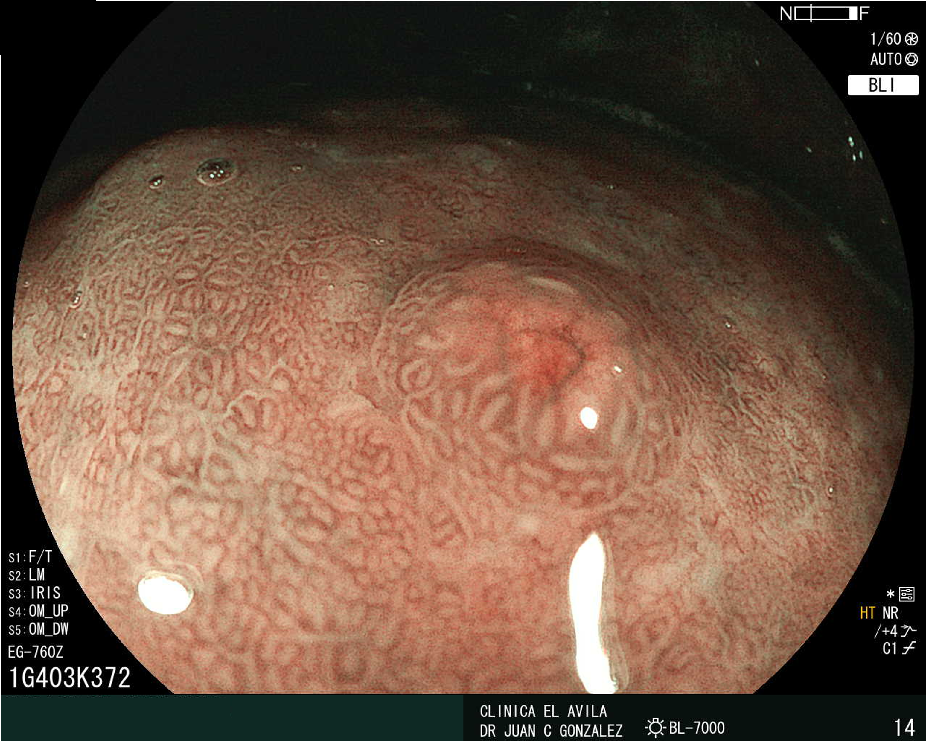
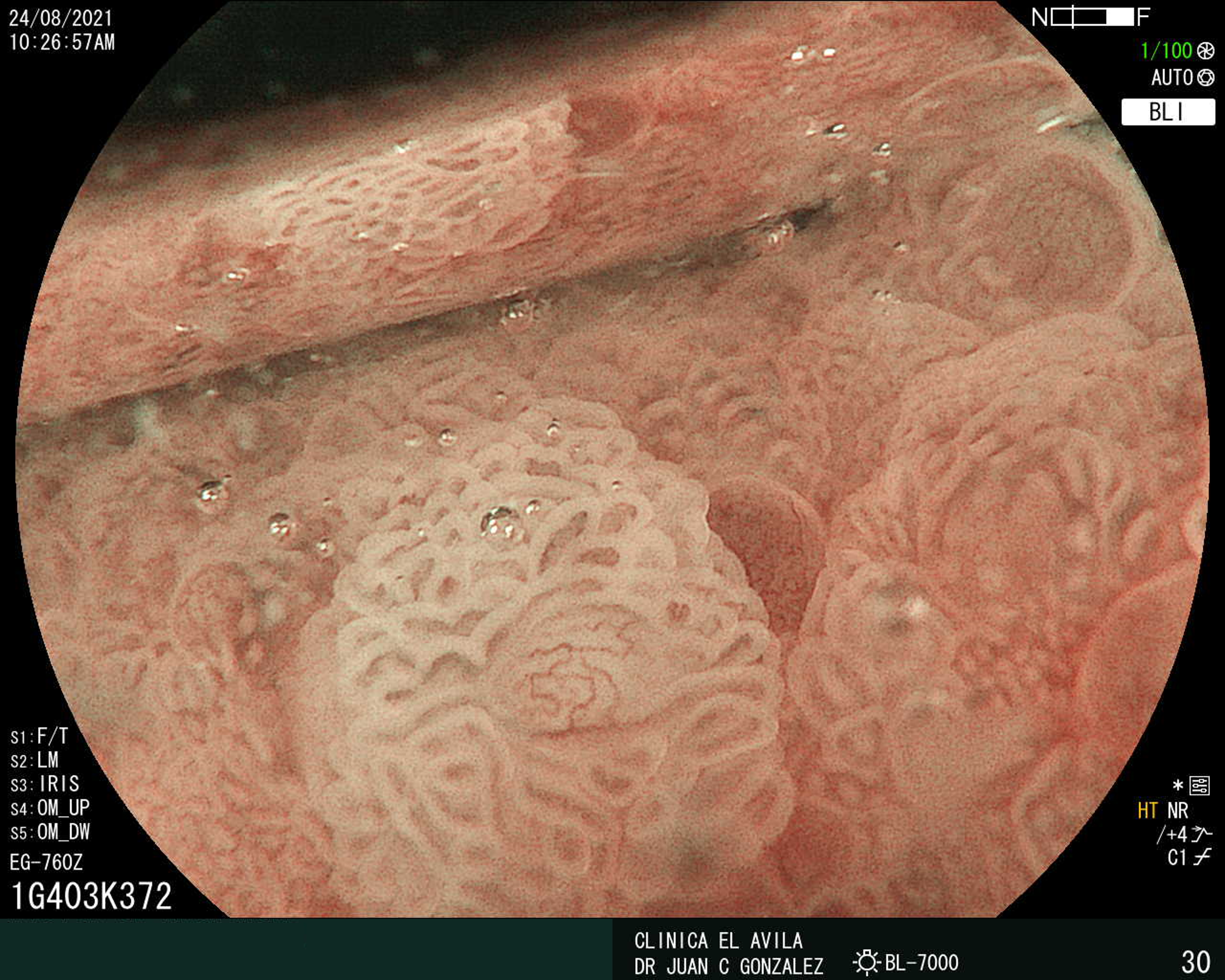
| Figura 1. Tumor Neuroendocrino Gástrico | Figura 2. Hiperplasia de Células Neuroendocrinas en estómago |
 |  |
 |  |  |
ISSN: 0016-3503 e-ISSN: 2477-975X


