Anemia en la enfermedad celíaca
Resumen
La anemia es una manifestación frecuente de la enfermedad celiaca en adultos. Por tanto, la sola presencia de anemia hace imperiosa la búsqueda activa de esta patología. La causa de la anemia en la enfermedad celiaca es multifactorial, la ferropénica es la más frecuente. El déficit de hierro puede explicarse por malabsorción, ingesta inadecuada, microsangrados intestinales y además por la descamación del epitelio intestinal. En general no responde a la suplementación oral con hierro, pero sí a la dieta libre de gluten. El déficit de ácido fólico es frecuente también ya que comparte sitio de absorción con el hierro. En pacientes que cumplen la dieta libre de gluten puede perpetuarse por el uso de harinas no suplementadas con el aporte mínimo requerido. Aunque la afectación ileal que provoque déficit de vitamina B 12 es poco prevalente, su déficit no es despreciable en estos pacientes y se asocia a alteraciones en la secreción ácida y al sobrecrecimiento bacteriano que puede estar asociado. La anemia inflamatoria también es una causa para considerar como en cualquier enfermedad crónica. Cursa con inhibición de la eritropoyetina por el estado proinflamatorio y aumento de los niveles de ferritina y hepcidina. En todos los casos el cumplimiento de la dieta libre de gluten es fundamental. En un paciente celiaco que cumple la dieta y persiste con anemia luego de un período considerable, debe considerarse de acuerdo con el contexto, que se trate de enfermedad celíaca refractaria o que exista una complicación. De lo contrario, obliga a buscar diferenciales independientes de la enfermedad celiaca.
Palabras clave
Texto completo:
PDFReferencias
Ludvigsson JF, Leffler DA, Bai JC, et al. The Oslo definitions forcoeliac disease and related terms. Gut 2013; 62(1): 43–52.
Bai JC, Ciacci C, Corazza GR, et al. Guías Mundiales de la Organización Mundial de Gastroenterología – Enfermedad celíaca. Última versión 2016. (Internet)
Al-Toma A, Volta U, Auricchio R, et al. European Society for the Study of CoeliacDisease (ESsCD) guideline for coeliac diseaseand other gluten-related disorders. United European GastroenterolJ. 2019; 7(5): 583–613.
Rubio-Tapia A, Hill ID, Kelly CP, et al.ACG Clinical Guidelines: Diagnosis and Management ofCeliac Disease. Am J Gastroenterol 2013; 108: 656–76.
Pinto-Sanchez MI,BercikP,Verdu EF, et al. Extraintestinal manifestations of celiac disease. Dig. Dis.2015, 33, 147–54.
Caio G, Volta U, Sapone A, et al. Celiac disease: a comprehensive current review. BMC Medicine 2019; 17: 142.
Baydoun A, Maakaron JE, Halawi H, et al. Hematological manifestations of celiacdisease. Scand. J. Gastroenterol. 2012; 47: 1401–11.
Catal F, Topal E, Ermistekin H, et al. The hematologicmanifestations of pediatric celiac disease at the time of diagnosis and efficiency of gluten free diet. Turk. J. Med. Sci. 2015; 45: 663–7.
Berry N, Basha J, Varma N, et al. Anemia in celiac disease is multifactorial in etiology: A prospective study from India. JGH Open 2018; 2: 196–200.
Mahadev, S.; Laszkowska, M.; Sundstrom, J.; Björkholm, M.; Lebwohl, B.; Green, P.H.R.; Ludvigsson, J.F.Prevalence of celiac disease in patients with iron deficiency anemia—A systematic review and meta-analysis.Gastroenterology 2018, 155, 374–82.
Wierdsma NJ, van Bokhorst-de van derScheuren MA, Berkenpas M, et al. Vitamin and mineral deficiencies are highly prevalent in newly diagnosed celiac disease patients. Nutrients2013, 5, 3975–92.
Abu Daya H, Lebwohl B, Lewis SK, et al. Celiac diseasepatients presenting with anemia have more severe disease thanthose presenting with diarrhea. ClinGastroenterolHepatol 2013;11: 1472-7.
Baydoun A, Maakaron JE, Halawi H, et al. Hematological manifestations of celiacdisease. Scand. J. Gastroenterol. 2012; 47: 1401–11.
Mooney PD, Kurien M, Evans KE, et al. Clinical and immunologic features of ultra-short celiac disease. Gastroenterology 2016; 150: 1125–34.
Doyev R, Cohen S, Ben-Tov A, et al. Ultra-short celiac disease is a distinct and milder phenotype of the disease in children. Dig. Dis. Sci. 2019; 64: 167–72.
Repo M, Lindfors K, Mäki M, et al. Anemia and Iron Deficiency in Children with Potential CeliacDisease. J. Pediatr. Gastroenterol. Nutr. 2017; 64: 56–62.
Murray JA, McLachlan S, Adams PC, et al. Association between celiac diseaseand iron deficiency in Caucasians, but not non-Caucasians. ClinGastroenterolHepatol 2013; 11: 808-14.
Kosnai I, Kuitunen P, Siimes MA. Iron deficiency in children withcoeliac disease on treatment with gluten-free diet. Role of intestinalblood loss. Arch Dis Child 1979; 54: 375-8.
Fine KD. The prevalence of occult gastrointestinal bleeding inceliac sprue. N Engl J Med 1996; 334: 1163-7.
Russo M, Elichalt M, Vázquez D, et al. Fortificación de harina de trigo con ácido fólico y hierro en Uruguay; implicancias en la nutrición. Rev ChilNutr 2014; 41 (4): 399-403.
Freeman HJ. Iron deficiency anemia in celiac disease. World J Gastroenterol 2015; 21(31): 9233-8.
Ivanovski P, Nikolić D, Dimitrijević N, et al. Erythrocytictransglutaminase inhibition hemolysis at presentationof celiac disease. World J Gastroenterol 2010; 16: 5647-50.
Dawson AM, Holdsworth CD, Pitcher CS. Sideroblastic anaemiain adult coeliac disease. Gut 1964; 5: 304-8.
Hendrickx GF, Somers K, Vandenplas Y. Lane-Hamiltonsyndrome: case report and review of the literature. Eur J Pediatr 2011; 170: 1597-1602.
Taytard J, Nathan N, de Blic J, et al. New insights into pediatricidiopathic pulmonary hemosiderosis: the French RespiRare(®)cohort. Orphanet J Rare Dis 2013; 8: 161.
Vaucher P, Druais PL, Waldvogel S, et al. Effect of ironsupplementation on fatigue in nonanemic menstruating womenwith low ferritin: a randomized controlled trial. CMAJ 2012; 184: 1247-54.
Illing AC, Shawki A, Cunningham CL, et al. Substrateprofile and metal-ion selectivity of human divalent metal-iontransporter-1. J BiolChem 2012; 287: 30485-96.
Iolascon A, De Falco L. Mutations in the gene encoding DMT1:clinical presentation and treatment. SeminHematol 2009; 46:358-70.
Cherukuri S, Potla R, Sarkar J, etal.Unexpected role of ceruloplasmin in intestinal iron absorption. Cell Metab 2005; 2: 309-19.
Finch C. Regulators of iron balance in humans. Blood 1994; 84: 1697-102.
Sham RL, Phatak PD, West C, et al. Autosomal dominant hereditary hemochromatosis associated with a novel ferroportin mutation and unique clinical features. Blood Cells Mol Dis 2005; 34: 157-61.
Gulec S, Anderson GJ, Collins JF. Mechanistic and regulatoryaspects of intestinal iron absorption. Am J PhysiolGastrointestLiverPhysiol 2014; 307: G397-G409.
Liu Q, Davidoff O, Niss K, et al. Hypoxia-inducible factorregulates hepcidin via erythropoietin-induced erythropoiesis. J Clin Invest 2012; 122: 4635-44.
Barisani D, Parafioriti A, Bardella MT, et al. Adaptive changesof duodenal iron transport proteins in celiac disease. Physiol Genomics 2004; 17: 316-25.
Sharma N, Begum J, Eksteen B, et al. Differential ferritin expressionis associated with iron deficiency in coeliac disease. EurJGastroenterolHepatol 2009; 21: 794-804.
Korman SH. Pica as a presenting symptom in childhood celiacdisease. Am J ClinNutr 1990; 51: 139-41.
Balaban DV, Popp A, Beata A, et al. Diagnostic accuracy of red blood cell distributionwidth-to-lymphocyte ratio for celiac disease. Rev. Romana Med. Lab. 2018; 26: 45–50.
Johnson-Wimbley TD, Graham DY. Diagnosis and management of iron deficiency anemia in the 21st
century. Adv. Gastroenterol. 2011; 4: 177–84.
Jericho H, SansottaN,Guandalini S. Extraintestinal Manifestations of Celiac Disease: Efectiveness of theGluten-Free Diet. J. Pediatr. Gastroenterol. Nutr. 2017; 65: 75–9.
Saez LR, Alvarez DF, Martinez IP, et al. Refractory iron-deficiency anemiaand gluten intolerance — response to gluten-free diet. Rev EspEnferm Dig. 2011; 103: 349–54.
Hopper AD, Leeds JS, Hurlstone DP, et al. Are lowergastrointestinal investigations necessary in patients with coeliac disease? Eur. J. Gastroenterol. Hepatol. 2005; 17: 617–21.
Balaban DV,PoppA, Radu FI, et al. Hematologic Manifestations in Celiac Disease—APractical Review. Medicina 2019; 55 (373): 1-8.
Annibale B, Severi C, Chistolini A, et al. Efficacy ofgluten-free diet alone on recovery from iron deficiency anemia inadult celiac patients. Am J Gastroenterol 2001; 96: 132-7.
García Á, Lucendo A. Review: Nutritional and Dietary Aspects of Celiac Disease. Nutr. Clin. Pract, 2011; 26: 163–73.
Martín R, Nestares M, Díaz J, et al. Multifactorial Etiology of Anemia in Celiac Disease and Effect of Gluten-Free Diet: A Comprehensive Review. Nutrients, 2019: 2557.
Harper JW, Holleran SF, Ramakrishnan R, et al.Anemia in celiac disease is multifactorial in etiology. Am. J. Hematol. 2007; 82: 996–1000.
Bergamaschi G, Markopoulos K, Albertini R, et al. Anemia of chronic disease and defectiveerythropoetin production in patients with celiacdisease. Hematologica 2008; 93: 1785–91.
Berry N, Basha J, Varma N, et al. Anemia in celiac disease is multifactorial in etiology: A prospective study from India. JGH Open, 2018; 2(5): 196–200.
Basu A, Ray Y, Bowmik P, et al. Rare association of coeliac disease with aplastic anemia. Report of a case from India. Indian J. Hematol. Blood Transfus. 2014; 30: 208–11.
Chatterjee S, Dey PK, Roy P, et al. Celiac disease with pure red cell aplasia: An unusual hematologicassociation in pediatric age group. Indian J. Hematol. Blood Transfus. 2014; 30: 383–5
DOI: http://dx.doi.org/10.61155/gen.v75i2.559
IMÁGENES GEN
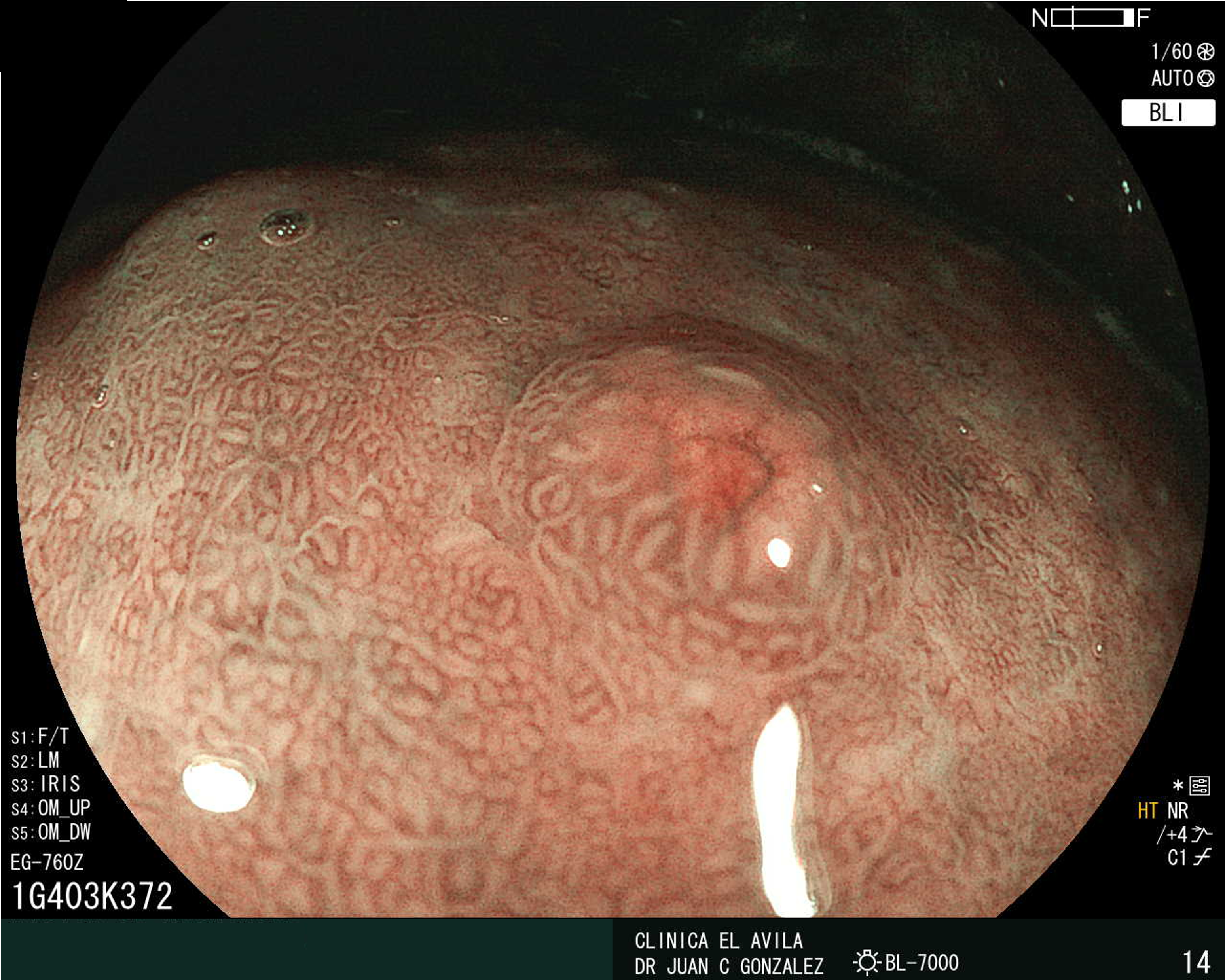
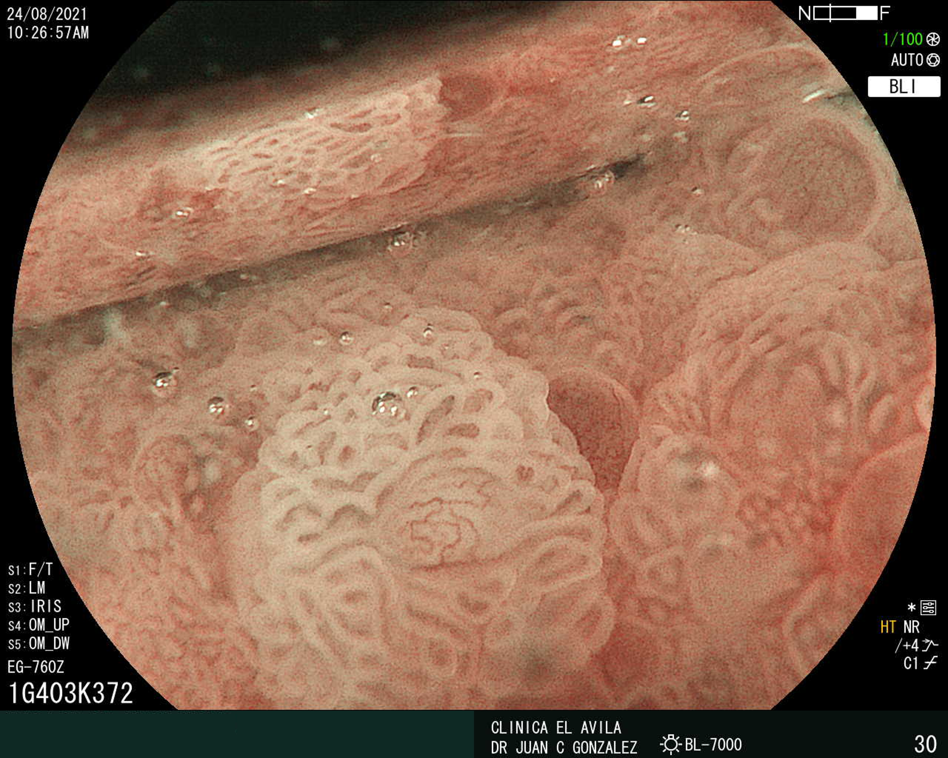
| Figura 1. Tumor Neuroendocrino Gástrico | Figura 2. Hiperplasia de Células Neuroendocrinas en estómago |
 |  |
 |  |  |
ISSN: 0016-3503 e-ISSN: 2477-975X


