Enfermedad Celíaca. Enfoque anatomopatológico de la enfermedad
Resumen
Palabras clave
Texto completo:
PDFReferencias
Sleisenger And Fordtran. Enfermedades Gastrointestinales y Hepáticas. Editorial Panamericana. 7ma edición. 2006. 92:1935-1962.
Trier JS. Diagnosis of celiac sprue. Gastroenterology. 1998;115(1):211-6.
Ludvigsson J, Leffler D, Bai JC, et al. The Oslo definitions for coeliac disease-related terms. Gut 2013 Jan;62(1):43-52. doi: 10.1136/gutjnl-2011-301346.
Ferguson A, Arranz E, O’Mahony S. Clinical and pathological spectrum of coeliac disease— active, silent, latent, potential. Gut 1993; 34: 150–1.
Green PH. The many faces of celiac disease: clinical presentation of celiac disease in the adult population. Gastroenterology 2005; 128 (Suppl 1): S74–S78.
Bain Julio, Fried Michael, Corazza Gino et al. Enfermedad Celíaca. Guías Mundiales de la Organización Mundial de Gastroenterología. 2012. chrome-extension://efaidnbmnnnibpcajpcglclefindmkaj/https://www.worldgastroenterology.org/UserFiles/file/guidelines/celiac-disease-spanish-2013.pdf
Masachs M, Casellas F, Malagelada JR. Enfermedad inflamatoria intestinal en pacientes celiacos. Rev Esp Enf Dig 2007; 99: 446-50.
Bai J, Zeballos E, Fried M. Corazza G,Schuppan D, Farthing M et al. World Gastroenterology Organisation Practice Guidelines: Enfermedad celíaca. En http://www.intramed.net/userfiles/file/ enfermedad_celiaca.pdf (Revisado en línea el 06 de julio de 2011).
Freeman HJ, Chopra A, Clandinin MT, Thomson AB. Recent advances in celiac disease. World J Gastroenterol 2011; 17: 2259-72.
Marsh MN. Gluten major histocompatibility complex, and the small intestine. Gastroenterology 1992; 102: 330–54. Guías Mundiales de la WGO (versión larga) 17 © World Gastroenterology Organization, 2012
Díaz Solángel, Dib Jr Jacobo. Enfermedad celíaca. Gen 2008;.62(3).
Green PH, Jabri B. Coeliac disease. Lancet 2003; 362: 383–91.
Perera DR, Weinstein WM, Rubin CE. Small intestinal biopsy. Hum Pathol. 1975; 6: 157- 217. http://dx.doi.org/10.1016/S0046-8177(75)80176-6 216)
Brenes-Pino F, Herrera A. La biopsia intestinal y su interpretación. Resultados preliminares en Costa Rica. En Rodrigo L y Pena AS, editores. Enfermedad celíaca y sensibilidad al gluten no celíaca. Barcelona, España: Omnia Science; 2013. p. 203-218
Grupo de trabajo del Protocolo para el diagnóstico precoz de la enfermedad celíaca. Protocolo para el diagnóstico precoz de la enfermedad celíaca. Ministerio de Sanidad, Servicios Sociales e Igualdad. Servicio de Evaluación del Servicio Canario de la Salud (SESCS). España, 2018. http://portal.guiasalud.es/contenidos/iframes/documentos/opbe/201805/SESCS_2018_Protocolo_diag_precoz_EC.pdf Ministerio de Sanidad y Consumo España Diagnóstico precoz de la enfermedad celíaca, Madrid
José Alfredo Murillo Saviano William Piedra Carvajal Daniela Sequeira Calderón Ellen Sylvie Sánchez Más Daniel Sandoval Loría. Generalidades de Enfermedad Celiaca y abordaje diagnóstico. TEMA 9 -2019. Revista Clínica de la Escuela de Medicina UCR-HSJD. 2019; 9(2): 64-69. ISSN-2215 2741.
Julio C. Bai, Michael Fried, Gino Roberto Corazza, Detlef Schuppan et al. Enfermedad Celiaca. Guías Mundiales de la Organización Mundial de Gastroenterología. 2012
Brizuela LO, Villadóniga RC, Santisteban SHN, et al. Enfermedad celíaca en el adulto. Un reto en el nuevo milenio. Mul Med 2020;24(4):949-968.
Rubio A, Hill I, Ciarán K, Calderwood A Murray J. Diagnosis and Management of Celiac Disease. Am J Gastroenterol 2013; 108: 656–676; doi:10.1038/ajg.2013.79.https://gi.org/guideline/diagnosis-and-management-of-celiac-disease,
AGA Institute. AGA Institute Medical Position Statement on the Diagnosis and Management of Celiac Disease. Gastroenterol. 2006; 131: 1977-80.
Wolf J Petroff D Richter T, et al. Validation of Antibody-Based Strategies for Diagnosis of Pediatric Celiac Disease without Biopsy. Gastroenterology 2017; 153:410–419. https://www.ncbi.nlm.nih.gov/pubmed/28461188.
Mangiavillano B, Parma B, Brambillasca MF, Albarello L, Barera G, Mariani A et al. Diagnostic bulb biopsies in celiac disease. Gastrointest Endosc. 2009; 69: 388-9.
Bonamico M, Thanasi E, Mariani P, Nenna R, Luparia RPL, Barbera C et al. Duodenal Bulb Biopsies in Celiac Disease: A multicenter Study. J Ped Gastroenterol and Nutr. 2008; 47:618-22)
Isabel Frahm. Cómo manejar las muestras anatomopatológicas para obtener buenos resultados (histológicos, inmunohistoquímicos, moleculares y genéticos). E d i t o r i a l. Revista Argentina de Mastología 2017; 36 (30).
Babbin BA, Crawford K, Sitaraman SV. Malabsorpton work-up: utlity of small bowel biopsy. Clin Gastroenterol Hepatol. 2006; 4: 1193-8. http://dx.doi.org/10.1016/j.cgh.2006.07.022
Brenes F. Observación personal.
Marsh MN. Gluten major histocompatibility complex, and the small intestine. Gastroenterology 1992; 102: 330–54.
Green PH, Jabri B. Coeliac disease. Lancet 2003; 362: 383–91.
Marsh MN, Crowe PT. Morphology of the mucosal lesion in gluten sensitivity. Baillire's Clinical Gastroenterology 1995; 2: 273-293.
Cerf-Bensussan N, Cerf M, Grand G.Gut intraepithelial lymphocytes and gastrointestinal diseases. Curr Opin Gastroenterology 1993; 9: 953-961.
Russell GJ, Winter HS, Fox VL, Bhan AK. Lymphocytes bearing the gamma-delta T receptor in normal human intestine and celiac disease. Hum Pathol 1991; 22: 690-694.
Ensari A. Gluten-sensitive enteropathy (celiac disease): controversies in diagnosis and classilication. Arch Pathol Lab Med 2010;134(6):826-36.
Srivastava A. Oberhuber versus Marsh: much ado about nothing? Gastroenterol Hepatol Bed Bench. 2015;8(4):244-6.
Ensari A. Coeliac disease: to classily ar not to classily - that is the question! Gastroenterol Hepatol Bed Bench 2016; 9(2):73-4. doi: 10.1043/1543-2165-134.6.826-
Oberhuber, G. Histopathology of celiac disease. Biomed Pharmacother 2000; 54: 368-72.
Corazza GR, Villanacci V, Zambelli C, Milione M, Luinetti O, Vindigni C, et al. Comparison of the interobserver reproducibility with different histologic criteria used in celiac disease. Clin Gastroenterol Hepatol 2007; 5: 838-43.
Di Sabatino A, Corazza G. Coeliac disease. Lancet 2009; 373: 1480-94.
Rubio-Tapia A, Hill I, Kelly C, Calderwood A, Murray J. ACG Clinical Guidelines: Diagnosis and Management of Celiac Disease. Am J Gastroenterol 2013; 108: 656-76.
Husby S, Koletzko S, Korponay-Szabo I, Mearin ML, Phillips A, Shamir R, et al. European Society for Pediatric Gastroenterology, Hepatology, and Nutrition guidelines for the diagnosis of coeliac disease. J Pediatr Gastroenterol Nutr 2012; 54: 136-60.
Walker M, Murray J, Ronkainen J, Aro P, Storskrubb T, D'Amato M, et al. Detection of celiac disease and lymphocytic enteropathy by parallel serology and histopathology in a population-based study. Gastroenterology 2010; 139: 112-9.
Aziz I, Evans K, Hopper A, Smillie D, Sanders D. A prospective study into the aetiology of lymphocytic duodenosis. Aliment Pharmacol Ther 2010; 32: 1392-7.
Moscoso JF, Quera PR. Update on celiac disease. Rev Med Chil. 2016;144(2):211-21. doi: 10 .4067/S0034-98872016000200010
Srivastava A. Oberhuber versus Marsh: much ado about nothing? Gastroenterol Hepatol Bed Bench 2015;8(4):244-6.
Kelly CP, Bai JC, Liu E, Leffler DA. Advances in diagnosis and management of celiac disease. Gastroenterology. 2015;148(6):1175-86. doi: 10.1053/j.gastro.2015.01.044.
Stoven SA, Choung RS, Rubio-Tapia A, Absah I, Lam-Himlin DM, Harris LA, Ngamruengphong S, Vazquez Roque MI, Wu TT, Murray JA. Analysis of biopsies from duodenal bulbs of all endoscopy patients increases detection of abnarmalities but has a minimal effect on diagnosis of celiac disease. Clin Gastroenterol Hepatol . 2016;14(11):1582-1588. doi: 10.1016/j.cgh.2016.02.026.
Campos Ruiz A, Urtasun Arlegui L, Marra-López Valenciano C. Sprue-like enteropathy linked to olmesartan. Rev Esp Enferm Dig 2016;108(5):292-3. doi: 10.17235/reed.2016.4140/2015.
laniro G, Gasbarrini A, Cammarota G. Olmesartan¬associated sprue-like enteropathy: know your enemy. Scand J Gastroenterol 2016;51(7):891. doi: 10.3109/00365521.2016.1153139.
Arguedas Lázaro Y, Santolaria Piedralita S. Enfermedad celíaca. Medicine. 2016;12(4):168-77. doi: 10.1016/j.med.2016.02.010
Husby S, Koletzko S, Karponay-Szabó IR, Mearin ML, Phillips A, Shamir R, Troncone R, Giersiepen K, Branski D, Catassi C, Lelgeman M, Miiki M, Ribes-Koninckx C, Ventura A, Zimmer KP; ESPGHAN Warking Group on Coeliac Disease Diagnosis; ESPGHAN Gastroenterology Committee; European Society for Pediatric Gastroenterology, Hepatology, and Nutrition. European Society for Pediatric Gastroenterology, Hepatology, and Nutrition guidelines for the diagnosis of coeliac disease. J Pediatr Gastroenterol Nutr. 2012;54(1):136-60. doi: 10.1097/MPG.0b013e31821a23d0
Marsh MN. Coeliac disease, mucosal change and IEL: doing what counts the best. Gastroenterol Hepatol Bed Bench 2016;9(1):1-5.
Biagi F, Vattiato C, Burrone M, Schiepatti A, Agazzi S, Maiarano G, Luinetti O, Alvisi C, Klersy C, Corazza GR. Is a detailed grading of villous atrophy necessary lar the diagnosis 01 enteropathy? J Clin Pathol 2016;69(12):1051-1054. doi: 10.1136/jclinpath-2016-203711.
Kelly CP, Bai JC, Liu E, Leffler DA. Advances in diagnosis and management of celiac disease. Gastroenterology. 2015 May; 148(6):1175-86. doi: 10.1053/j.gastro.2015.01.044
Oberhuber G, Granditsch G, Vogelsang H. The histopathology of coeliac disease: tme for a standardized report scheme for pathologists. Eur J Gastroenterol Hepatol. 1999; 11: 1185-94. http://dx.doi.org/10.1097/00042737-199910000-00019
Goldstein NS, Underhill J. Morphologic features suggestve of gluten sensitvity in architecturally normal duodenal biopsy specimens. Am J Clin Pathol. 2001; 116: 63-71. http://dx.doi.org/10.1309/5PRJ-CM0U-6KLD-6KCM
Goldstein NS. Proximal small-bowel mucosal villous intraepithelial lymphocytes. Histopathol. 2004; 44: 199-205. http://dx.doi.org/10.1111/j.1365-2559.2004.01775.x
Jarvinen TT, Collin P, Rasmussen M, Kyronpalo S, Maki M, Partanen J et al. Villous tp intraepithelial lymphocytes as markers of early-stage coeliac disease. Scand J Gastroenterol. 2004; 39: 428-33. http://dx.doi.org/10.1080/00365520310008773
Biagi F, Luinet O, Campanella J, Klersy C, Zambelli C, Villanacci V et al. Intraepithelial lymphocytes in the villous tp:do they indicate potental coeliac disease? J Clin Pathol. 2004; 57: 835-9.http://dx.doi.org/10.1136/jcp.2003.013607
Brown I, Mino-Kenudson M, Deshpande V, Lauwers GY. Intraepithelial lymphocytosis in architecturally preserved proximal small intestnal mucosa: an increasing diagnostic problem with a wide diferental diagnosis. Arch Pathol Lab Med. 2006; 130: 1020-5.
DOI: http://dx.doi.org/10.61155/gen.v76i4.459
IMÁGENES GEN
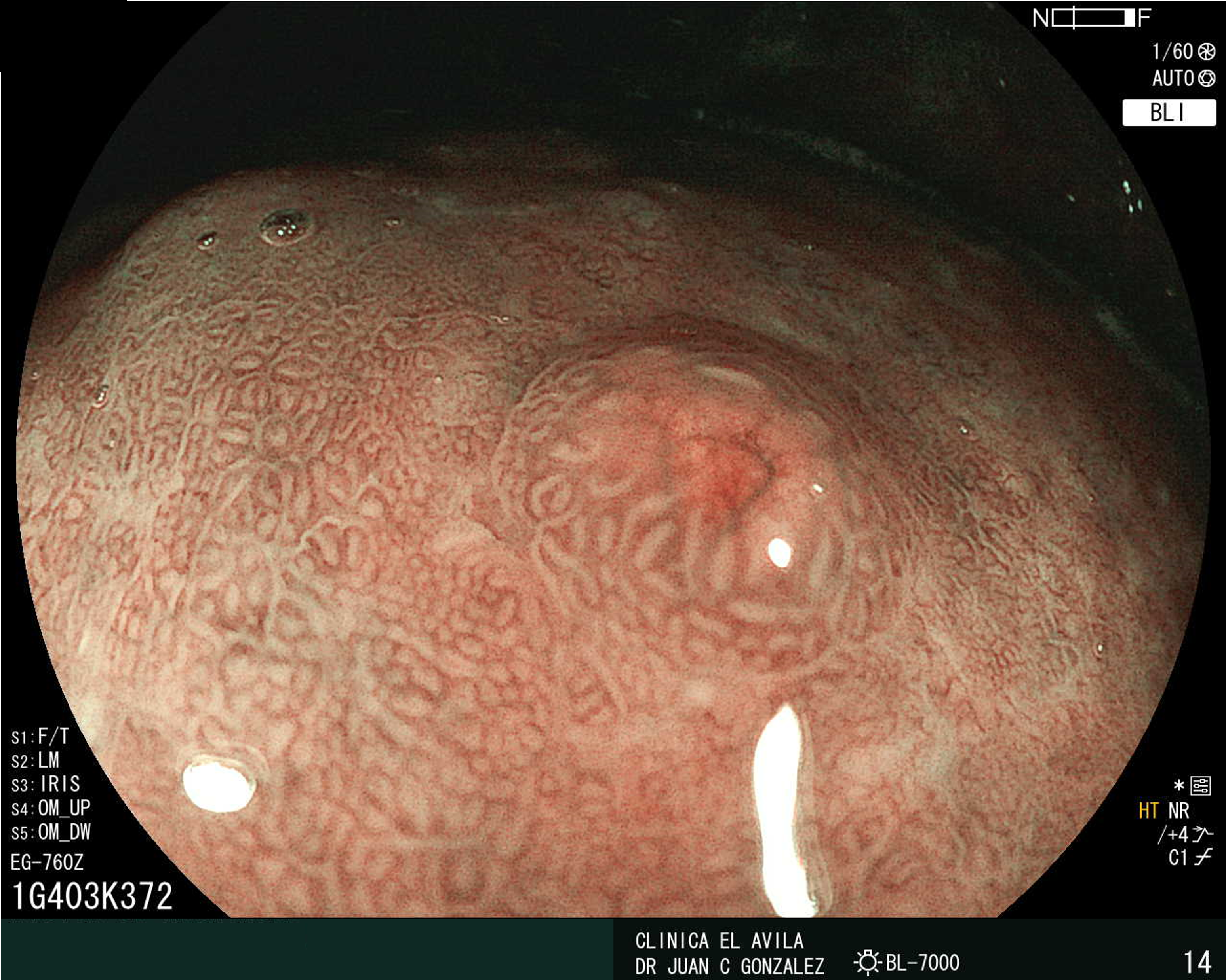
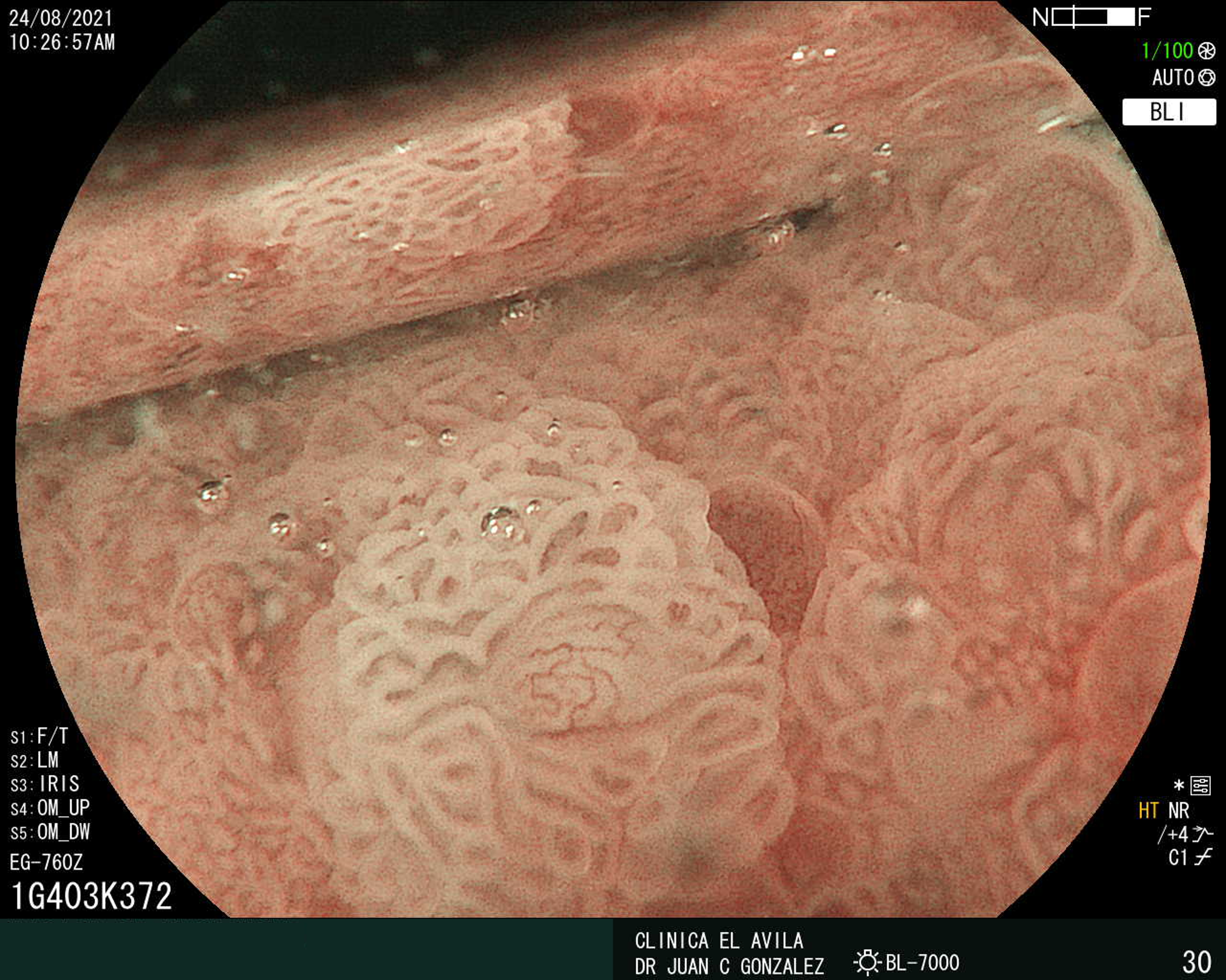
| Figura 1. Tumor Neuroendocrino Gástrico | Figura 2. Hiperplasia de Células Neuroendocrinas en estómago |
 |  |
 |  |  |
ISSN: 0016-3503 e-ISSN: 2477-975X


