DETECCIÓN DEL CÁNCER PRECOZ DEL ESÓFAGO, ESTÓMAGO Y COLON MEDIANTE LA CROMOENDOSCOPIA VIRTUAL MÁS MAGNIFICACIÓN ENDOSCÓPICA
Resumen
Mundialmente el carcinoma del tracto gastrointestinal es el líder como causa de muerte global por cáncer. Su prevención se basa en la detección precoz endoscópica del cáncer potencialmente curable. El diagnóstico precoz permite la resección quirúrgica ó endoscópica curativa.
La endoscopia moderna con la cromoendoscopia virtual ó digital y magnificación, mejora la visualización del patrón de superficie mucosal y el patrón vascular; y ha logrado instalarse como método alternativo potencial para la detección del cáncer precoz del esófago, estómago y colo-rectal.
El sistema de cromoendoscopia virtual FICE (Flexible Spectral Imaging Color Enhacement) viene demostrando un incremento en el rendimiento diagnóstico, mediante una mejor caracterización de las lesiones neoplásicas del tubo digestivo, con alta sensibilidad, especificidad y seguridad diagnóstica.
Presentamos nuestra experiencia con el sistema FICE 4450 de cromoendoscopia virtual + magnificación endoscópica, en el diagnóstico precoz del cáncer de esófago, estómago en 712 pacientes, y colo-rectal en 600 pacientes mayores de 40 años de edad, examinados entre septiembre 2012 a diciembre 2016 y su correlación histopatológica. Los resultados obtenidos indican la alta eficacia del sistema FICE 4450, en la identificación y caracterización de las lesiones, permitiendo el diagnóstico precoz del cáncer gastrointestinal.
Palabras claves: Endoscopia moderna con imagen mejorada. Sistema FICE (Flexible Imagen de Color Espectral Mejorada). Cromoendoscopia virtual y magnificación. Detección del cáncer precoz del esófago, estómago y colo-rectal.
Palabras clave
Texto completo:
PDFReferencias
Berr F, Oyama T, Ponchon T and Yahagi N: Early Neoplasias of the Gastrointestinal Tract. Endoscopic Diagnosis and therapeutic decisions. Springer (New York) 2014:3-33
Internacional Agency for Research on Cancer. WHO. World Cancer Report 2014
Globocan 2012 v1.1. Cancer Incidence and Mortality Worldwide: IARC Cancer Base nº 11. Lyon, France; 2014
Coda S and Thillainayagam A: State of the art in advanced endoscopic image for the detection and evaluation of dysplasia and early cancer of the gastrointestinal tract. Clin Exp Gastroenterol 2014;7: 133-150
Negreanu L, Preda C, Ionescu D and Ferechide D. Progress in digestive endoscopy: Flexible Spectral Imaging Colour Enhancement (FICE)-technical review. J Med Life 2015; 8 (4): 416-422
Osawa H, Yamamoto H, Miura Y, Ajibe H, Shinhata H and Yosizawa M: Diagnosis of depressed-type early gastric cancer using small caliber endoscopy with flexible spectral imaging color enhancement. Dig Endosc 2012; 24 (4): 231-236
Osawa H, Yamamoto H, Miura Y, et al: Diagnosis and extent of early gastric cancer using flexible spectral imaging color enhancement. World J Gastrointest Endosc 2012; 4 (8): 356-361
Hellier M and Williams J. The burden of gastrointestinal disease: implications for the provision of care in the UK. Gut 2007; 56 (2) 165-166
Aparcero M, González J, Argotte R, et al: Neoplasia gástrica maligna precoz y avanzado. Análisis endoscópico y correlación patológica. GEN 1986; 40: 4-9
Rey J, Kiesslich R and Hoffman A: New aspects of modern endoscopy. World J Gastrointest Endosc 2014; 6 (8): 334-344
Lambert R, Saito H and Saito Y. High-resolution endoscopy and early gastrointestinal cancer…dawn in the East. Endoscopy 2007; 39 (3) 232-237
Osawa H and Yamamoto H: Present and future status of flexible spectral imaging color enhancement and blue laser imaging technology. Dig Endosc 2014; 26 (1): 105-115
Li Y, Shen L, Yu H, Luo H and Yu J; Fujinon intelligent color enhancement for the diagnosis of early esophageal squamous cell carcinoma and precancerous lesion. Turk J Gastroenterol 2014; 25 (4): 365-369
Inoue H, Honda T, Nagai K, et al: Ultrahigh magnification endoscopic observation of carcinoma in situ of the esophagus. Dig Endosc 1997; 9:16-18
Kumagai Y, Inoue H, Nagai K, Kawano T and Takeshita K: Magnifying endoscopic, stereoscopic microscopy and the microvascular architecture of superficial esophageal carcinoma. Endoscopy 2002; 34:369-375
Misumi A, Harad K, Murakami A, et al: Role of lugol dye endoscopy in the diagnosis of early esophageal cancer. Endoscopy 1990; 22 (1): 12-16
Takenaka R, Kawahara Y, Okeda H, Hori K, Inoue M and Kawano S: Narrow-band imaging provides reliable screening for esophageal malignancy in patients with head and neck cancers. The American Journal of Gastroenterology 2009; 104 (12): 2942-2948
Fujita R. Early Cancer of the Gastrointestinal Tract. Springer, 2006
Takuto K. Histopathology 2007; 51: 733-742
Ishihara R, Inoue T, Uedo N, Yamamoto S, Kawada N and Tsujii Y: Significance each of narrow-band imaging finding in diagnosing squamous mucosal high-grade neoplasia of esophagus. J Gastroenterol Hepatol 2010; 25 (8): 1410-5
Participants in the Paris Workshop: The Paris endoscopic clasification of superficial neoplastic lesions: esophagus, stomach and colon. Gastrointest Endosc 2003; 58 (6): 53-522
Sano T, Kobori O and Muto T: “Lymph node metastasis from early gastric cancer: endoscopic resection of tumour”. The British Journal of Surgery 1992; 79 (3): 241-244
Gotoda T, Yanagisawa A, Sasako M, Ono H, Nakanishi Y and Shimoda T: Incidence of lymph node metastasis from early gastric cancer: estimation with a large number of cases at two large centers. Gastric cancer 2000; 3: 219-25
Kikuste I, Marques R, Monteiro M, et al: Systematic review of the diagnosis of gastric premalignant conditions and neoplasia with high resolution endoscopic technologies. Scand J Gastroenterol 2013; 48 (10): 1108-1117
Yao K, Anagnostopoulos GK and Ragunath K: Magnifying endoscopy for diagnosing and delineating early gastric cancer. Endoscopy 2009; 41: 462-467
Yao K, Iwashita A, Matsui T, Nambu M, Tanabe H and Nagahama T: White opaque substance within superficial-elevated gastric neoplasia as visaulizaed by magnification endoscopy (ME) with narrow-band imaging (NBI): A new useful maker for discriminating adenoma from carcinoma. Endoscopy 2007; 39: A16.G
Yoshizawa M, Osawa H, Yamamoto H, et al: Diagnosis of elevated-type early gastric cancer by the optimal band imaging system. Gastrointest Endosc 2009; 69 (1): 19-27
Kudo S, Tamura S, Nakajima T, Yamano H, Kusaka H and Watanabe H: Diagnosis of colorectal tumorous lesions by magnifying endoscopy. Gastrointest Endosc 1996; 44 (1): 8-14
Tanaka S and Sano Y: Aim to unify the narrow band imaging (NBI) magnifying classification for colorectal tumors: current status in Japan from a summary of the consensus in the 79th Annual Meeting of the Japan Gastroenterological Endoscopy Society. Dig Endosc 2011; 23 (1): 131-9
Yoshida N, Naito Y, Kugai M, Inoue K, Uchiyama K and Takagi T : Efficacy of magnifying endoscopy with flexible spectral imaging color enhancement in the diagnosis of colorectal tumors. J Gastroenterol 2011; 46: 65-72
Shimoda T, Ikegami M, Fujisaki J, Matsui T, Aizawa S and Ishikawa E: Early colorectal carcinoma with special reference to its development de-novo. Cancer 1989; 64 (5): 1138-46
Tateishi Y, Nakanishi Y, Taniguchi H, Tadakazu S and Satoshi U: Pathological prognostic factors predicting lymph node metastasis in submucosal invasive (T1) colorectal carcinoma. Modern Pathology 2010; 23: 1068-1072
Yasuda K, Inomata M, Shiromizu A, Shiraishi N, Higashi H and Kitano S: Risk factors for occult lymph node metastasis of colorectal cancer invading the submucosa and indications for endoscopic mucosal resection. Dis Colon Rectum 2007; 50 (9): 1370-6
Westwood D, Alexakis N, Connor S, et al: Transparent cap-assisted colonoscopy versus standard adult colonoscopy: a systematic review and meta-analysis. Dis Colon Rectum 2012; 55: 218-225
DOI: http://dx.doi.org/10.61155/gen.v71i4.373
IMÁGENES GEN
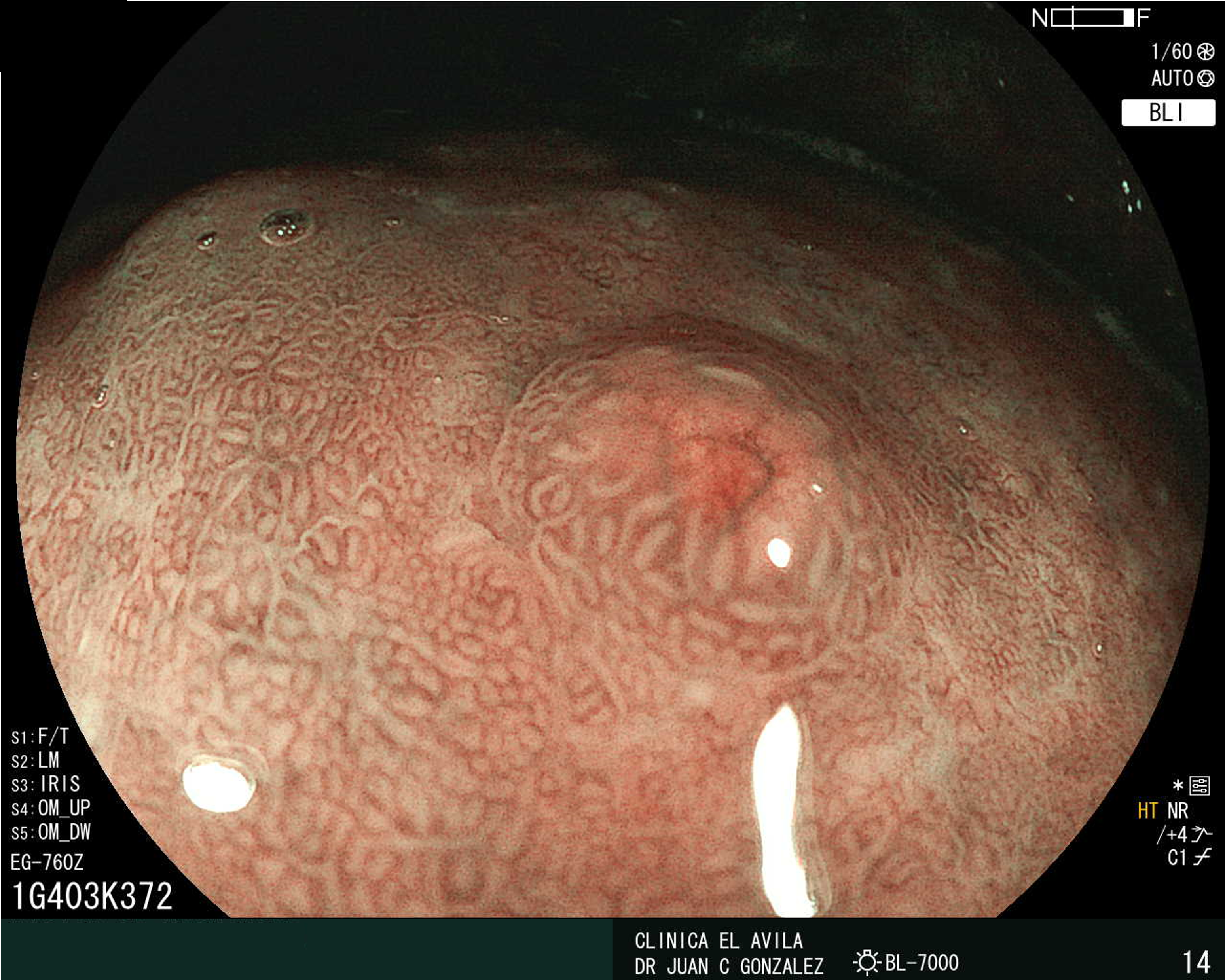
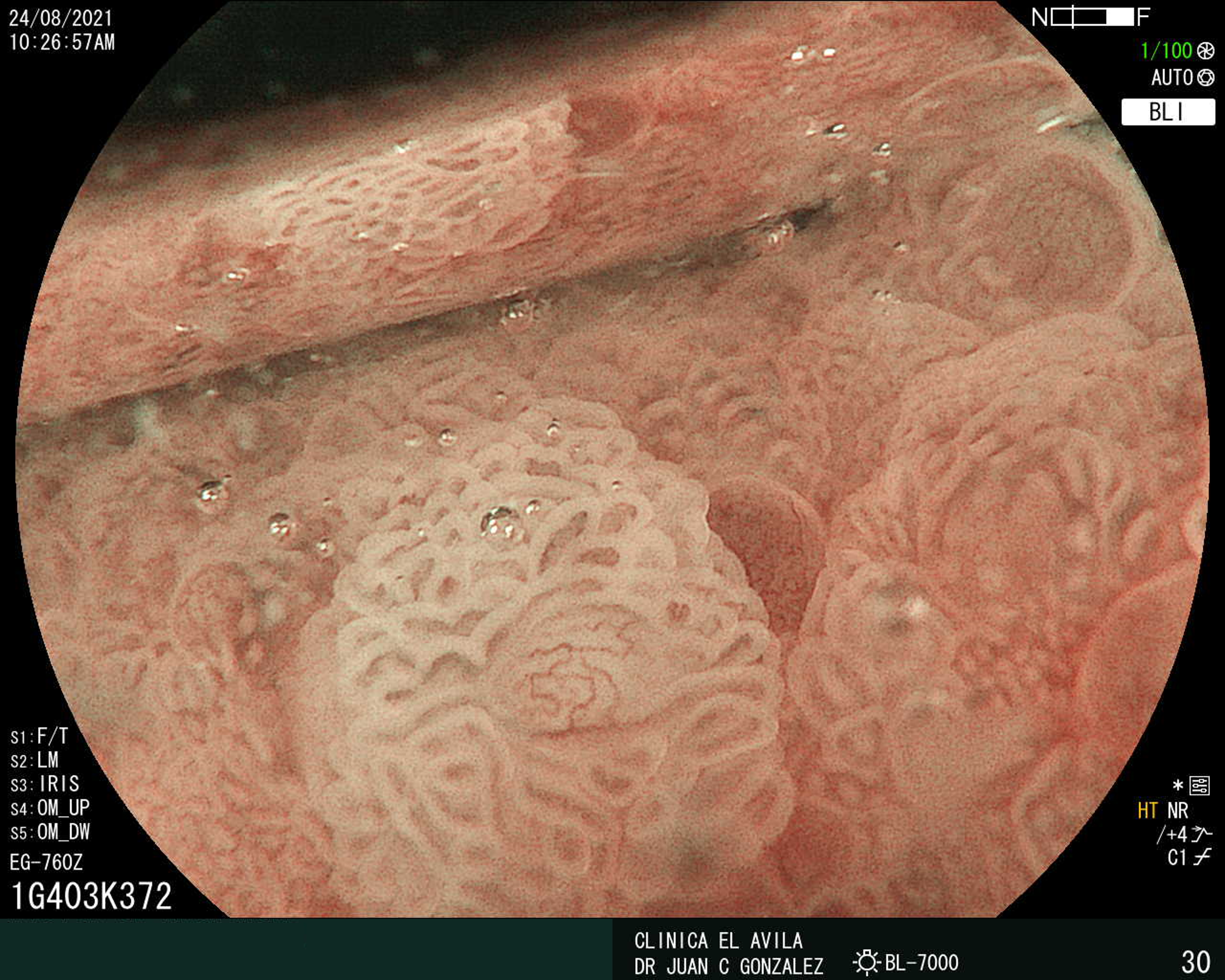
| Figura 1. Tumor Neuroendocrino Gástrico | Figura 2. Hiperplasia de Células Neuroendocrinas en estómago |
 |  |
 |  |  |
ISSN: 0016-3503 e-ISSN: 2477-975X


 ESCUCHAR RESUMEN DEL ARTICULO